Cervical Spine X Ray / Normal flexion and extension cervical spine x-rays | Image ... - The density should be appropriate with soft tissues and bony structures well visualized.
Cervical Spine X Ray / Normal flexion and extension cervical spine x-rays | Image ... - The density should be appropriate with soft tissues and bony structures well visualized.. Fracture of the ring of c1 can allow the anterior portion of the ring to migrate anteriorly with respect to the dens. It shows cervical vertebra one through five. The cervical vertebrae make up the neck. It assists in planning and assessing prior to the surgery taking place. You want to see the entire cervical spine so that you can make sure that there is not an injury there.
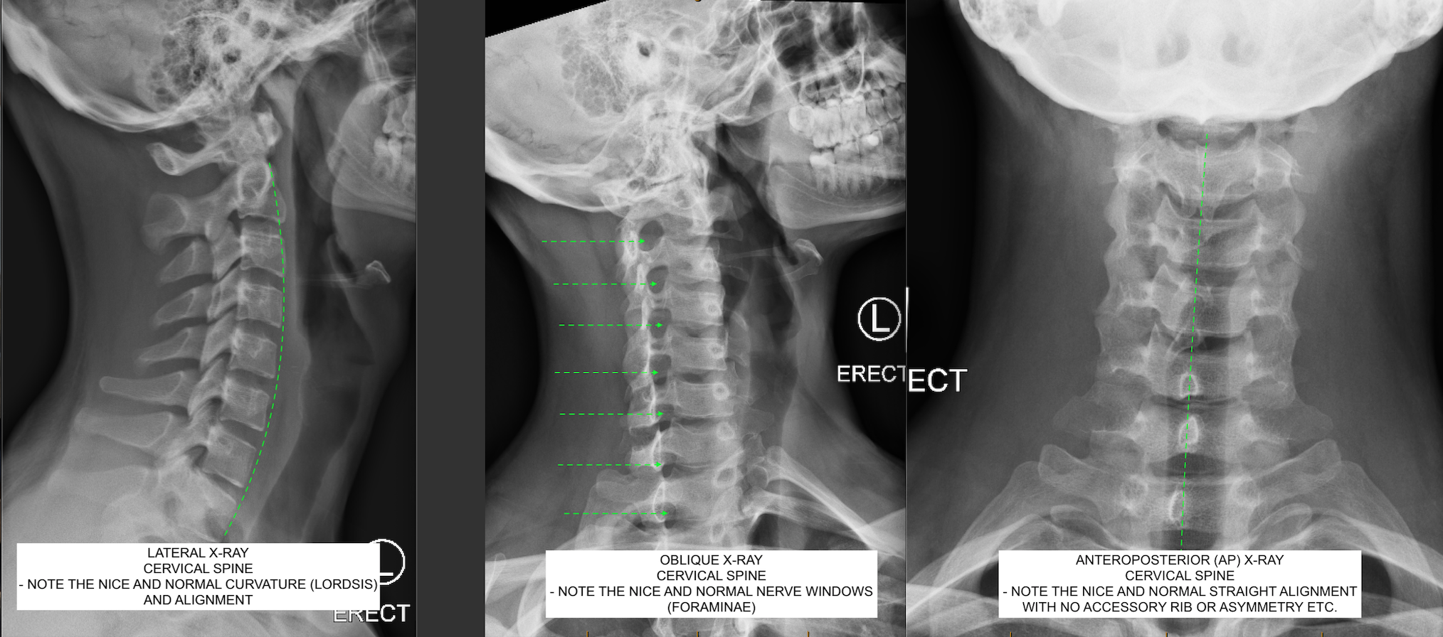
You require all three views (lateral, ap and odontoid/open mouth view) for an adequate study. The first method is done routinely on ambulatory patients in the outpatient department. You need to see either a neurosurgeon or an orthopedist doctor. An important advantage is the opportunity to get this diagnosis in the framework. Make sure you can see all 7 cervical spinous process.
Normal Cervical Spine Xray | Bone and Spine from i2.wp.com The first method is done routinely on ambulatory patients in the outpatient department. Radiology schools radiology student medical student radiology imaging medical imaging radiologic technology cervical spondylosis spinal stenosis medical anatomy. Fracture of the ring of c1 can allow the anterior portion of the ring to migrate anteriorly with respect to the dens. They may be taken to find injuries or diseases that affect the discs or joints in your spine. An important advantage is the opportunity to get this diagnosis in the framework. You need to see either a neurosurgeon or an orthopedist doctor. One can look at the health of the discs, uncovertebral joints, facets, nerve root foramen, alignment, spinal canal diameter, and even the. You require all three views (lateral, ap and odontoid/open mouth view) for an adequate study.
You want to see the entire cervical spine so that you can make sure that there is not an injury there.
So what are we looking at? When reading any radiograph the clinician should establish a process or order they follow each time. You require all three views (lateral, ap and odontoid/open mouth view) for an adequate study. The standard three views taken are the Several injury patterns can widen this space. You might have a herniated disc in your cervical area. The first method is done routinely on ambulatory patients in the outpatient department. Lying down method is done in cases of injury or patients who cannot stand. Normal cervical spine radiographs in a young adult. Cervical spine radiographic series contains 3 views. Ct scan of the cervical spine. Left without, right with annotation. You want to see the entire cervical spine so that you can make sure that there is not an injury there.
You need to see either a neurosurgeon or an orthopedist doctor. Left without, right with annotation. Look for alignment of four parallel vertical columns that follow a slightly lordotic curve without any step offs. You might have a herniated disc in your cervical area. You want to see the entire cervical spine so that you can make sure that there is not an injury there.
C.N.S. Neurosurgery | Normal Cervical Spine X-ray from www.cnsneurosurgery.com.au The thoracic vertebrae comprise the chest section and there is low radiation exposure. Cervical spine radiographic series contains 3 views. The cervical vertebrae make up the neck. He pointed out my yoinked neck and was shocked that i wasn't in a serious amount of neck pain. An important advantage is the opportunity to get this diagnosis in the framework. One can look at the health of the discs, uncovertebral joints, facets, nerve root foramen, alignment, spinal canal diameter, and even the. Normal cervical spine radiographs in a young adult. The spine is divided into several sections.
Most spinal injuries occur at the junctions of the spine.
The standard three views taken are the This post is real simple. Radiology schools radiology student medical student radiology imaging medical imaging radiologic technology cervical spondylosis spinal stenosis medical anatomy. Most spinal injuries occur at the junctions of the spine. The cervical vertebrae make up the neck. One can look at the health of the discs, uncovertebral joints, facets, nerve root foramen, alignment, spinal canal diameter, and even the. You require all three views (lateral, ap and odontoid/open mouth view) for an adequate study. You want to see the entire cervical spine so that you can make sure that there is not an injury there. Fracture of the ring of c1 can allow the anterior portion of the ring to migrate anteriorly with respect to the dens. So what are we looking at? Make sure you can see all 7 cervical spinous process. Several injury patterns can widen this space. Normal cervical spine radiographs in a young adult.
Clearly can be seen that c2 (red outline) is moved forward with respect to c3 (blue outline). It assists in planning and assessing prior to the surgery taking place. They may be taken to find injuries or diseases that affect the discs or joints in your spine. Case contributed by dr andrew dixon ◉. This post is real simple.
Xray Image Cervical Spine View Shows Stock Photo 332802647 ... from image.shutterstock.com An important advantage is the opportunity to get this diagnosis in the framework. When reading any radiograph the clinician should establish a process or order they follow each time. They may be taken to find injuries or diseases that affect the discs or joints in your spine. Most spinal injuries occur at the junctions of the spine. Cervical spine pa or ap. One can look at the health of the discs, uncovertebral joints, facets, nerve root foramen, alignment, spinal canal diameter, and even the. Case contributed by dr andrew dixon ◉. You might have a herniated disc in your cervical area.
Look for alignment of four parallel vertical columns that follow a slightly lordotic curve without any step offs.
Fracture of the ring of c1 can allow the anterior portion of the ring to migrate anteriorly with respect to the dens. Several injury patterns can widen this space. Ct scan of the cervical spine. Clearly can be seen that c2 (red outline) is moved forward with respect to c3 (blue outline). Left without, right with annotation. The standard three views taken are the Look for alignment of four parallel vertical columns that follow a slightly lordotic curve without any step offs. You want to see the entire cervical spine so that you can make sure that there is not an injury there. Cervical spine radiographic series contains 3 views. Most spinal injuries occur at the junctions of the spine. When reading any radiograph the clinician should establish a process or order they follow each time. You might have a herniated disc in your cervical area. It shows cervical vertebra one through five.
You have just read the article entitled Cervical Spine X Ray / Normal flexion and extension cervical spine x-rays | Image ... - The density should be appropriate with soft tissues and bony structures well visualized.. You can also bookmark this page with the URL : https://mod-cuek.blogspot.com/2021/07/cervical-spine-x-ray-normal-flexion-and.html
Share Awesome
Belum ada Komentar untuk "Cervical Spine X Ray / Normal flexion and extension cervical spine x-rays | Image ... - The density should be appropriate with soft tissues and bony structures well visualized."

Belum ada Komentar untuk "Cervical Spine X Ray / Normal flexion and extension cervical spine x-rays | Image ... - The density should be appropriate with soft tissues and bony structures well visualized."
Posting Komentar